Search
Search Results
-
Detection of Cell Wall-Anchoring Machinery by Immunogold-Labeling Thin-Section Electron Microscopy
Cell wall anchoring of surface proteins and pili in Gram-positive bacteria is mediated by sortase – a highly conserved transpeptidase enzyme. Early...
-
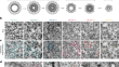
Immunogold Labeling of Milk Proteins at Transmission Electron Microscopy
Milk intended for human consumption is subjected to technological treatments to ensure its safety and storage stability. These treatments deeply...
-
Characterization of Extracellular Vesicles by Transmission Electron Microscopy and Immunolabeling Electron Microscopy
Transmission electron microscopy (TEM) is currently the only method that enables the observation of extracellular vesicles (EVs) at a nanometer...
-
Immunogold for Protein Location in Chromaffin Cells
The localization and density of any plasma membrane or intracellular protein in chromaffin cells are prerequisites for those studies designed to...
-
High-Pressure Freezing and Freeze Substitution for Transmission Electron Microscopy Imaging and Immunogold-Labeling
Electron microscopy enables the unbiased imaging of organelles and cellular structures at nano-meter scale resolution. The combination of...
-
NPC Structure in Model Organisms: Transmission Electron Microscopy and Immunogold Labeling Using High-Pressure Freezing/Freeze Substitution of Yeast , Worms, and Plants
The nuclear pore complex (NPC) is a large elaborate structure embedded within the nuclear envelope, and intimately linked to the cytoskeleton,...
-
Scanning Electron Microscopy (SEM) and Immuno-SEM of Nuclear Pore Complexes from Amphibian Oocytes, Mammalian Cell Cultures, Yeast, and Plants
Scanning electron microscopy (SEM)Scanning electron microscopy (SEM) can be used to image nuclear pore complex (NPC) surface structure Structures of...
-
Imaging Cytoskeleton Components by Electron Microscopy
The cytoskeleton is a complex of detergent-insoluble components of the cytoplasm playing critical roles in cell motility, shape generation, and...
-
Assessing CB1 Expression in the Brain by Immunohistochemical Methods: Light, Confocal, and Electron Microscopy
Conventional techniques to reveal the neuroanatomical distribution of type 1 cannabinoid receptor (CB1) in the brain, at the cellular and subcellular...
-
Genetically encoded barcodes for correlative volume electron microscopy
While genetically encoded reporters are common for fluorescence microscopy, equivalent multiplexable gene reporters for electron microscopy (EM) are...
-
Localization of Mitochondrial Nucleoids by Transmission Electron Microscopy Using the Transgenic Expression of the Mitochondrial Helicase Twinkle and APEX2
Reminiscent of their evolutionary origin, mitochondria contain their own genome (mtDNA) compacted into the mitochondrial chromosome or nucleoid...
-
Studying Plant ER-PM Contact Site Localized Proteins Using Microscopy
As in most eukaryotic cells, the plant endoplasmic reticulum (ER) network is physically linked to the plasma membrane (PM), forming ER-PM contact...
-
Electron Tomography of Cryo-Fixed and Resin-Embedded Samples
Room-temperature electron tomography of sections prepared from high-pressure frozen, freeze-substituted and resin-embedded cells is a powerful method...
-
Localizing Proteins on Single Trafficking Organelles in 3D with Semisynthetic Gold Labeling and Platinum Replica Electron Microscopy
The three-dimensional structures of organelles can be visualized at high resolutions using electron microscopy and tomography. Combining genetically...
-
Transmission Electron Microscopy and Tomography on Plasma Membrane Sheets to Study Secretory Docking
To study the formation and the architecture of exocytotic site, we generated plasma membrane (PM) sheets on electron microscopy grids to visualize...
-
Cryo-electron tomography on focused ion beam lamellae transforms structural cell biology
Cryogenic electron microscopy and data processing enable the determination of structures of isolated macromolecules to near-atomic resolution....

-
Imaging the inner structure of chromosomes: contribution of focused ion beam/scanning electron microscopy to chromosome research
Visualization of the chromosome ultrastructure has revealed new insights into its structural and functional properties. The use of new methods for...

-
Methods and Molecular Tools for Studying Endocytosis in Plants---an Overview
Proteins of the endocytosis machinery in plants, such as clathrin and adaptor proteins, were isolated and characterized using combinations of...
-
Targeting, Localisation and Identification in Cryo-ET
One of the main benefits of cryo-electron tomography (ET) is direct visualisation of the sample itself, without the need for stains or fixation. A...
-
Electron Tomography and Immunogold Labeling as Tools to Analyze De Novo Assembly of Plant Cell Walls
High-resolution imaging of the membranous intermediates and cytoskeletal arrays involved in the assembly of a new cell wall during plant cytokinesis...