Search
Search Results
-
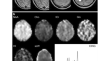
Microstructural and Metabolic Changes in Normal Aging Human Brain Studied with Combined Whole-Brain MR Spectroscopic Imaging and Quantitative MR Imaging
PurposeThis study aimed to detect age-related brain metabolic and microstructural changes in healthy human brains by the use of whole-brain proton...
-
AI-assisted Segmentation Tool for Brain Tumor MR Image Analysis
TumorPrism3D software was developed to segment brain tumors with a straightforward and user-friendly graphical interface applied to two- and...

-
MR Template-Based Individual Brain PET Volumes-of-Interest Generation Neither Using MR nor Using Spatial Normalization
For more anatomically precise quantitation of mouse brain PET, spatial normalization (SN) of PET onto MR template and subsequent template...

-
Rate of abnormalities in quantitative MR neuroimaging of persons with chronic traumatic brain injury
BackgroundMild traumatic brain injury (mTBI) can result in lasting brain damage that is often too subtle to detect by qualitative visual inspection...

-
Pediatric brain aneurysms: a review of 1458 brain MR angiograms
PurposeTo evaluate clinical and imaging characteristics of pediatric brain aneurysms.
Materials and MethodsA retrospective review of 1458 MR...

-
MR-based radiomics predictive modelling of EGFR mutation and HER2 overexpression in metastatic brain adenocarcinoma: a two-centre study
ObjectivesMagnetic resonance (MR)-based radiomics features of brain metastases are utilised to predict epidermal growth factor receptor (EGFR)...

-
Impact of brain segmentation methods on regional metabolism quantification in 18F-FDG PET/MR analysis
BackgroundAccurate analysis of quantitative PET data plays a crucial role in studying small, specific brain structures. The integration of PET and...

-
RU-Net: skull strip** in rat brain MR images after ischemic stroke with rat U-Net
BackgroundExperimental ischemic stroke models play a fundamental role in interpreting the mechanism of cerebral ischemia and appraising the...

-
Development and performance of SIAT bPET: a high-resolution and high-sensitivity MR-compatible brain PET scanner using dual-ended readout detectors
PurposePositron emission tomography/magnetic resonance imaging (PET/MRI) is a powerful tool for brain imaging, but the spatial resolution of the PET...

-
An active learning approach to train a deep learning algorithm for tumor segmentation from brain MR images
PurposeThis study focuses on assessing the performance of active learning techniques to train a brain MRI glioma segmentation model.
Methods ...
-
An improved 3D-UNet-based brain hippocampus segmentation model based on MR images
ObjectiveAccurate delineation of the hippocampal region via magnetic resonance imaging (MRI) is crucial for the prevention and early diagnosis of...

-
Simultaneous high-resolution whole-brain MR spectroscopy and [18F]FDG PET for temporal lobe epilepsy
PurposePrecise lateralizing the epileptogenic zone in patients with drug-resistant mesial temporal lobe epilepsy (mTLE) remains challenging,...

-
PET/MR: Functional and Molecular Imaging of Neurological Diseases and Neurosciences
This book aims to summarize the research progress of integrated PET/MR brain function and molecular imaging, and more importantly, clinical...
-
The added value of relative amide proton transfer (rAPT) to advanced multiparametric MR imaging for brain glioma characterization
BackgroundDifferentiation between the grades of brain gliomas is a crucial step in the management of patients. The gold standard technique for...

-
An Effective Approach to Improve the Automatic Segmentation and Classification Accuracy of Brain Metastasis by Combining Multi-phase Delay Enhanced MR Images
The objective of this study is to analyse the diffusion rule of the contrast media in multi-phase delayed enhanced magnetic resonance (MR) T1 images...

-
Evaluation of T2W FLAIR MR image quality using artificial intelligence image reconstruction techniques in the pediatric brain
BackgroundArtificial intelligence (AI) reconstruction techniques have the potential to improve image quality and decrease imaging time. However,...

-
In vivo demonstration of globotriaosylceramide brain accumulation in Fabry Disease using MR Relaxometry
PurposeHow to measure brain globotriaosylceramide (Gb3) accumulation in Fabry Disease (FD) patients in-vivo is still an open challenge. The objective...

-
Fetal brain MR angiography at 1.5 T: a feasible study
PurposeThe use of magnetic resonance angiography (MRA) for assessing CNS fetal vasculature has been limited. The aim of this study was to determine...

-
Application of a Modified Combinational Approach to Brain Tumor Detection in MR Images
For many years, brain tumor detection has been one of the most essential and competitive issues for medical researchers. Many methods have been...

-
Quantitative study of the changes in brain white matter before and after radiotherapy by applying multi-sequence MR radiomics
PurposeTo analyse the changes in brain white matter before and after radiotherapy (RT) by applying multisequence MR radiomics features and to...
