Search
Search Results
-
Morphological analysis of descending tracts in mouse spinal cord using tissue clearing, tissue expansion and tiling light sheet microscopy techniques
Descending tracts carry motor signals from the brain to spinal cord. However, few previous studies show the full view of the long tracts from a 3D...

-
Projective light-sheet microscopy with flexible parameter selection
Projection imaging accelerates volumetric interrogation in fluorescence microscopy, but for multi-cellular samples, the resulting images may lack...

-
Benchtop mesoSPIM: a next-generation open-source light-sheet microscope for cleared samples
In 2015, we launched the mesoSPIM initiative, an open-source project for making light-sheet microscopy of large cleared tissues more accessible....

-
descSPIM: an affordable and easy-to-build light-sheet microscope optimized for tissue clearing techniques
Despite widespread adoption of tissue clearing techniques in recent years, poor access to suitable light-sheet fluorescence microscopes remains a...

-
Minutes-timescale 3D isotropic imaging of entire organs at subcellular resolution by content-aware compressed-sensing light-sheet microscopy
Rapid 3D imaging of entire organs and organisms at cellular resolution is a recurring challenge in life science. Here we report on a computational...

-
Blazed oblique plane microscopy reveals scale-invariant inference of brain-wide population activity
Due to the size and opacity of vertebrate brains, it has until now been impossible to simultaneously record neuronal activity at cellular resolution...

-
Multi-immersion open-top light-sheet microscope for high-throughput imaging of cleared tissues
Recent advances in optical clearing and light-sheet microscopy have provided unprecedented access to structural and molecular information from intact...

-
Structure and assembly of the S-layer in C. difficile
Many bacteria and archaea possess a two-dimensional protein array, or S-layer, that covers the cell surface and plays crucial roles in cell...

-
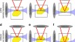
Cancellation of Bessel beam side lobes for high-contrast light sheet microscopy
An ideal illumination for light sheet fluorescence microscopy entails both a localized and a propagation invariant optical field. Bessel beams and...

-
Multidirectional digital scanned light-sheet microscopy enables uniform fluorescence excitation and contrast-enhanced imaging
Light-sheet fluorescence microscopy (LSFM) has emerged as a powerful method for rapid and optically efficient 3D microscopy. Initial LSFM designs...

-
Programmable RNA base editing with photoactivatable CRISPR-Cas13
CRISPR-Cas13 is widely used for programmable RNA interference, imaging, and editing. In this study, we develop a light-inducible Cas13 system called...

-
Imaging peripheral nerve micro-anatomy with MUSE, 2D and 3D approaches
Understanding peripheral nerve micro-anatomy can assist in the development of safe and effective neuromodulation devices. However, current approaches...

-
4-bit adhesion logic enables universal multicellular interface patterning
Multicellular systems, from bacterial biofilms to human organs, form interfaces (or boundaries) between different cell collectives to spatially...

-
EDTP enhances and protects the fluorescent signal of GFP in cleared and expanded tissues
Advanced 3D high-resolution imaging techniques are essential for investigating biological challenges, such as neural circuit analysis and tumor...

-
Self-assembly of a layered two-dimensional molecularly woven fabric
Fabrics—materials consisting of layers of woven fibres—are some of the most important materials in everyday life
1 . Previous nanoscale weaves2 –16 ...
-
Versatile whole-organ/body staining and imaging based on electrolyte-gel properties of biological tissues
Whole-organ/body three-dimensional (3D) staining and imaging have been enduring challenges in histology. By dissecting the complex physicochemical...

-
Myelin dysfunction drives amyloid-β deposition in models of Alzheimer’s disease
The incidence of Alzheimer’s disease (AD), the leading cause of dementia, increases rapidly with age, but why age constitutes the main risk factor is...

-
Automatic and adaptive heterogeneous refractive index compensation for light-sheet microscopy
Optical tissue clearing has revolutionized researchers’ ability to perform fluorescent measurements of molecules, cells, and structures within intact...
-
In-situ particle analysis with heterogeneous background: a machine learning approach
We propose a novel framework that combines state-of-the-art deep learning approaches with pre- and post-processing algorithms for particle detection...
-
SSPIM: a beam sha** toolbox for structured selective plane illumination microscopy
Selective plane illumination microscopy (SPIM) represents a preferred method in dynamic tissue imaging, because it combines high spatiotemporal...
