Search
Search Results
-
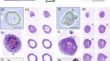
CohortFinder: an open-source tool for data-driven partitioning of digital pathology and imaging cohorts to yield robust machine-learning models
Batch effects (BEs) refer to systematic technical differences in data collection unrelated to biological variations whose noise is shown to...

-
Applications of artificial intelligence in the analysis of histopathology images of gliomas: a review
In recent years, the diagnosis of gliomas has become increasingly complex. Analysis of glioma histopathology images using artificial intelligence...

-
Deep learning-based virtual H& E staining from label-free autofluorescence lifetime images
Label-free autofluorescence lifetime is a unique feature of the inherent fluorescence signals emitted by natural fluorophores in biological samples....

-
In vivo organoid growth monitoring by stimulated Raman histology
Patient-derived tumor organoids have emerged as a crucial tool for assessing the efficacy of chemotherapy and conducting preclinical drug screenings....
-
Increased [18F]FDG uptake of radiation-induced giant cells: a single-cell study in lung cancer models
Positron emission tomography (PET), a cornerstone in cancer diagnosis and treatment monitoring, relies on the enhanced uptake of fluorodeoxyglucose ([

-
Emerging paradigms in microwave imaging technology for biomedical applications: unleashing the power of artificial intelligence
In recent years, microwave imaging (MWI) has emerged as a non-ionizing and cost-effective modality in healthcare, specifically within medical...

-
Macrophage PET imaging in mouse models of cardiovascular disease and cancer with an apolipoprotein-inspired radiotracer
Macrophages are key inflammatory mediators in many pathological conditions, including cardiovascular disease (CVD) and cancer, the leading causes of...

-
Personalized coronary and myocardial blood flow models incorporating CT perfusion imaging and synthetic vascular trees
Computational simulations of coronary artery blood flow, using anatomical models based on clinical imaging, are an emerging non-invasive tool for...

-
In vivo imaging using surface enhanced spatially offset raman spectroscopy (SESORS): balancing sampling frequency to improve overall image acquisition
In the field of optical imaging, the ability to image tumors at depth with high selectivity and specificity remains a challenge. Surface enhanced...

-
Nondestructive, longitudinal, 3D oxygen imaging of cells in a multi-well plate using pulse electron paramagnetic resonance imaging
The use of oxygen by cells is an essential aspect of cell metabolism and a reliable indicator of viable and functional cells. Here, we report partial...

-
Artificial intelligence unravels interpretable malignancy grades of prostate cancer on histology images
Malignancy grading of prostate cancer (PCa) is fundamental for risk stratification, patient counseling, and treatment decision-making. Deep learning...

-
Self-assembled peptide-dye nanostructures for in vivo tumor imaging and photodynamic toxicity
We report noncovalent assemblies of iRGD peptides and methylene blue dyes via electrostatic and hydrophobic stacking. These resulting nanomaterials...

-
Multimodal bioimaging across disciplines and scales: challenges, opportunities and breaking down barriers
Multimodal bioimaging is a broad term used to describe experimental workflows that employ two or more different imaging modalities. Such approaches...
-
Tantalum oxide nanoparticles as versatile and high-resolution X-ray contrast agent for intraductal image-guided ablative procedure in rodent models of breast cancer
There are limited options for primary prevention of breast cancer (BC). Experimental procedures to locally prevent BC have shown therapeutic efficacy...

-
A Raman topography imaging method toward assisting surgical tumor resection
Achieving complete tumor resection upon initial surgical intervention can lead to better patient outcomes by making adjuvant treatments more...

-
Imaging cancer metabolism using magnetic resonance
The challenge in clinical oncology is to select the most appropriate treatment for an individual patient. Transcriptome and metabolite profiling have...

-
Fast histological assessment of adipose tissue inflammation by label-free mid-infrared optoacoustic microscopy
Conventional histology, as well as immunohistochemistry or immunofluorescence, enables the study of morphological and phenotypical changes during...
