Search
Search Results
-
Visualizing Metal Distribution in Plants Using Synchrotron X-Ray Fluorescence Microscopy Techniques
Recent improvements in synchrotron-based X-ray fluorescence (SXRF) microscopy established it as an advanced analytical tool for analyzing 2D- and 3D...
-
Correlative single-cell hard X-ray computed tomography and X-ray fluorescence imaging
X-ray computed tomography (XCT) and X-ray fluorescence (XRF) imaging are two non-invasive imaging techniques to study cellular structures and...

-
Multielement Z-tag imaging by X-ray fluorescence microscopy for next-generation multiplex imaging
Rapid, highly multiplexed, nondestructive imaging that spans the molecular to the supra-cellular scale would be a powerful tool for tissue analysis....

-
X-ray fluorescence spectroscopy (XRF) for metallome analysis of herbarium specimens
Background“Herbarium X-ray Fluorescence (XRF) Ionomics” is a new quantitative approach for extracting the elemental concentrations from herbarium...

-
In situ label-free X-ray imaging for visualizing the localization of nanomedicines and subcellular architecture in intact single cells
Understanding the intracellular behaviors of nanomedicines and morphology variation of subcellular architecture impacted by nanomaterial–biology...

-
Small-Angle X-Ray Scattering for Macromolecular Complexes
Small angle X-ray scattering (SAXS) is a versatile technique that can provide unique insights in the solution structure of macromolecules and their...
-
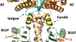
ATP-dependent conformational dynamics in a photoactivated adenylate cyclase revealed by fluorescence spectroscopy and small-angle X-ray scattering
Structural insights into the photoactivated adenylate cyclases can be used to develop new ways of controlling cellular cyclic adenosine monophosphate...
-
Gadolinium distribution in kidney tissue determined and quantified by micro synchrotron X-ray fluorescence
Aims of this study were to investigate gadolinium (Gd) in kidney tissue from a female patient with severe renal failure, who had a magnetic resonance...

-
Stimulated X-ray emission spectroscopy
We describe an emerging hard X-ray spectroscopy technique, stimulated X-ray emission spectroscopy (S-XES). S-XES has the potential to characterize...
-
Structure of cellulose in birch phloem fibres in tension wood: an X-ray nanodiffraction study
BackgroundTo gain a better understanding of bark layer structure and function, especially of the phloem fibres and their contribution to the posture...

-
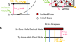
Monitoring Nuclease Activity by X-Ray Scattering Interferometry Using Gold Nanoparticle-Conjugated DNA
The biologically critical, exquisite specificity and efficiency of nucleases, such as those acting in DNA repair and replication, often emerge in the...
-
Multiscale X-ray phase-contrast CT unveils the evolution of bile infarct in obstructive biliary disease
Bile infarct is a pivotal characteristic of obstructive biliary disease, but its evolution during the disease progression remains unclear. Our...

-
An automated liquid jet for fluorescence dosimetry and microsecond radiolytic labeling of proteins
X-ray radiolytic labeling uses broadband X-rays for in situ hydroxyl radical labeling to map protein interactions and conformation. High flux density...

-
Penetration of foliar-applied Zn and its impact on apple plant nutrition status: in vivo evaluation by synchrotron-based X-ray fluorescence microscopy
The absorption of foliar fertilizer is a complex process and is poorly understood. The ability to visualize and quantify the pathway that elements...

-
X-ray Imaging of Root–Soil Interactions
Growing roots interact with soil, and its structure, across a range of spatial and temporal scales, and thus adapt to the local environment (Downie...
-
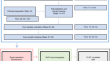
Automatic 3D cell segmentation of fruit parenchyma tissue from X-ray micro CT images using deep learning
BackgroundHigh quality 3D information of the microscopic plant tissue morphology—the spatial organization of cells and intercellular spaces in...

-
Neurotransmitter uptake of synaptic vesicles studied by X-ray diffraction
The size, polydispersity, and electron density profile of synaptic vesicles (SVs) can be studied by small-angle X-ray scattering (SAXS), i.e. by...

-
X-ray Ptychography Imaging of Human Chromosomes After Low-dose Irradiation
Studies of the structural and functional role of chromosomes in cytogenetics have spanned more than 10 decades. In this work, we take advantage of...

-
Sample preparation strategies for efficient correlation of 3D SIM and soft X-ray tomography data at cryogenic temperatures
3D correlative microscopy methods have revolutionized biomedical research, allowing the acquisition of multidimensional information to gain an...
-
Quantification of spatial metal accumulation patterns in Noccaea caerulescens by X-ray fluorescence image processing for genetic studies
BackgroundHyperaccumulation of trace elements is a rare trait among plants which is being investigated to advance our understanding of the regulation...
