Abstract
Transient receptor potential (TRP) non-selective cation channels constitute a superfamily, which contains 28 different genes. In mammals, this superfamily is divided into six subfamilies based on differences in amino acid sequence homology between the different gene products. Proteins within a subfamily aggregate to form heteromeric or homomeric tetrameric configurations. These different grou**s have very variable permeability ratios for calcium versus sodium ions. TRP expression is widely distributed in neuronal tissues, as well as a host of other tissues, including epithelial and endothelial cells. They are activated by environmental stresses that include tissue injury, changes in temperature, pH and osmolarity, as well as volatile chemicals, cytokines and plant compounds. Their activation induces, via intracellular calcium signalling, a host of responses, including stimulation of cell proliferation, migration, regulatory volume behaviour and the release of a host of cytokines. Their activation is greatly potentiated by phospholipase C (PLC) activation mediated by coupled GTP-binding proteins and tyrosine receptors. In addition to their importance in maintaining tissue homeostasis, some of these responses may involve various underlying diseases. Given the wealth of literature describing the multiple roles of TRP in physiology in a very wide range of different mammalian tissues, this review limits itself to the literature describing the multiple roles of TRP channels in different ocular tissues. Accordingly, their importance to the corneal, trabecular meshwork, lens, ciliary muscle, retinal, microglial and retinal pigment epithelial physiology and pathology is reviewed.
Introduction
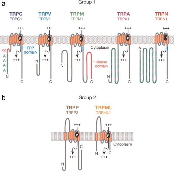
The founding member of the transient receptor potential (TRP) protein superfamily was first described in Drosophila. In some of these flies, prolonged exposure to light only induced transient photoreceptor depolarisation, whereas in the wild type the response was sustained. This difference was attributed to mutant trp gene expression. Mammalian TRP homologues have been identified over the past 30 years since the initial trp description in Drosophila. The members of this superfamily constitute a large and very different collection of proteins that are expressed in many tissues and cell types [1]. This superfamily is conserved throughout evolution, from nematodes to humans [2]. They form non-selective monovalent and divalent cation channels with very variable Ca2+/Na+ permeability ratios. Some members are even impermeable for Ca2+, whereas others are highly Ca2+ permeable relative to monovalent cations. TRPV5 and -6 exhibit a Ca2+/Na+ permeability ratio of greater than 100 [3]. This variability is unique by comparison with most other ion channel families. In those cases, the differences in permeation properties within a single family are for the most part very much smaller [4]. TRPs are distinguished from one another based on differences in their primary amino acid sequence rather than ligand affinity or selectivity. This system of classification is used because their properties are heterogeneous and their regulation is complex. The common feature of TRP channels is that they comprise six putative transmembrane spanning domains and a cationpermeable pore formed by a short hydrophobic region between transmembrane domains 5 and 6. They assemble themselves as homo-or heterotetramers to form cation channels (Figure 1). These channels are activated by a remarkable assemblage of very diverse stimuli.
Domain structure of the TRP superfamily. Five Group 1 TRP (TRPN is not expressed in mammals) (a) and two Group 2 TRP subfamilies (b) are listed. All subfamilies contain a six-transmembrane domain unit with a cation-permeable pore between domains I and II. Four such units are assembled as a hetero-or homotetramer to form TRP channels. Domain indications: ankyrin repeats (A), coiled-coil domain (cc), protein kinase domain (TRPM6/7 only), cation-permeable pore ( p), transmembrane (TM) domain, cation-permeable (+ + +), TRP domain (TRPC, TRPM only), large extracellular loop between TM I and II (TRPP, TRPML only). Adapted from Venkatachalam and Montell [5].
Genetic ablation studies in worms, flies and mice indicate that TRPs serve as sensors to elicit responses to a variety of stimuli, ranging from temperature, osmotic pressure, olfaction, taste, mechanical stress, light and injury-induced inflammation mediating pain. These channels are also activated by intracellular and extracellular messengers, as well as declines in the calcium content of intracellular Ca2+ stores [6]. The physiological importance of TRP expression is indicated by the finding that TRP mutations are linked to human diseases [7].
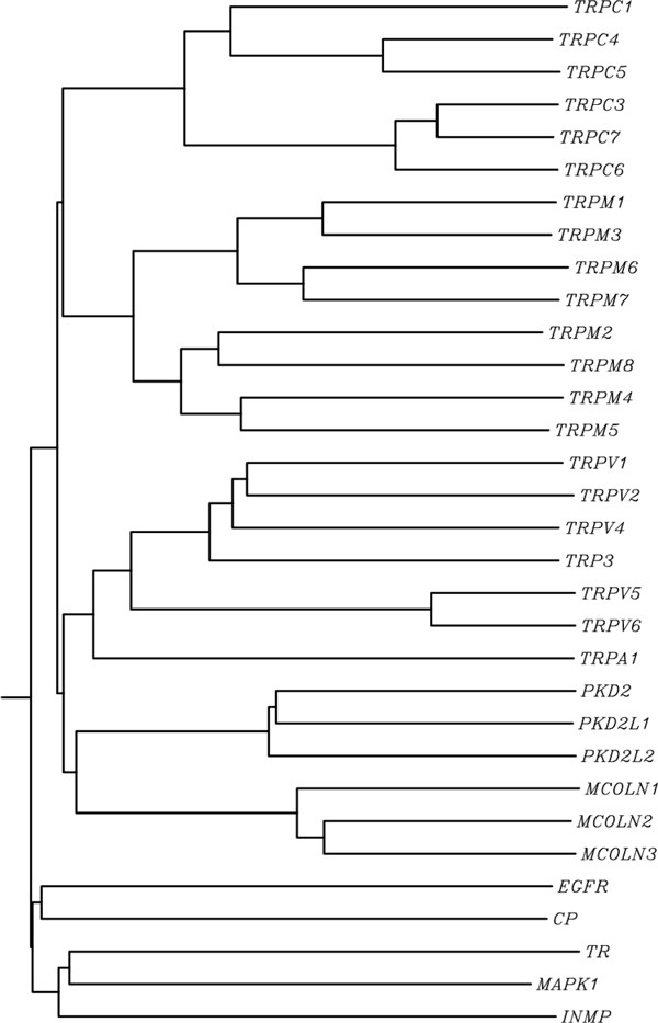
In humans, 27 different TRP genes are grouped into six different subfamilies based on their amino acid homologies (Table 1). The nomenclature of the TRP superfamily has been evolving for many years. Many genes and proteins within this superfamily have been referred to by multiple names, and in some cases two different genes belonging to different subfamilies have shared the same name. To overcome such confusion, a unified TRP superfamily nomenclature system was published in 2003, based on the TRPC ('Canonical'), TRPM ('Melastatin'), TRPV ('Vanilloid') subfamilies, with TRPA ('Ankyrin') being assigned subsequently [8]. These TRP subfamilies are designated as Group 1 TRPs. The nomenclature of the genes of Group 2 TRPs is based on the human phenotypes that the founding genes cause when mutated -- polycystic kinase disease (PKD) and mucolipidosis type IV (MCOLN, Mucolipin) -- while their encoded proteins have also been referred to as TRPP ('Polycystin') and TRPML ('Mucolipin'), respectively. Although the Group 2 proteins appear to fall into the same superfamily as Group 1 TRPs, Group 2 and Group 1 genes are only distally related (Figure 2).
The dendrogram of the TRP superfamily in humans. Random proteins bound to the plasma membrane (ie EGF receptor, EGFR), endoplasmic reticulum (ie calreticulin precursor, CP), mitochondria (thioredoxin reductase, TR), nuclear membrane (ie inner nuclear membrane protein, INMP) and a cystolic protein (ie mitogen-activated protein kinase 1, MAPK1) were selected to determine if PKD and MCOLN are evolutionarily related to TRP or the results of convergent evolution. Note that PKD and MCOLN belong to TRP superfamilies, since random genes extend from branches distinct from the TRP superfamily.
Group 1 TRPs share substantial sequence homology in the transmembrane domains. The TRPC subfamily consists of seven genes (ie TRPC1-7). TRPC2 is a pseudogene in humans. The TRPM subfamily comprises eight genes (ie TRPM 1-8), of which three encode channel-like proteins and five non-channel proteins. The TRPV subfamily contains six members (ie TRPV1-6). Recently, the TRPA subfamily was delineated and has one member [9]. TRPC, TRPV and TRPA channels contain ankyrin repeats in the intracellular N-terminal domain, whereas the TRPC and TRPM subfamilies possess a proline-rich 'TRP domain' in the region of the C terminal near the putative transmembrane segment. TRPM6 and -7 have a protein kinase domain in the C terminal.
The PKD and MCOLN gene branches encoding TRPP and TRPML channel proteins, respectively, belong to Group 2 TRPs. They are distally related to Group 1, as they contain limited sequence homology to Group 1 TRPs, although such homology is still greater than that exhibited by random genes with Group 1 TRPs (Figure 2). TRPP proteins share 25 per cent amino acid sequence similarity to TRPC3 and TRPC6 over a region including transmembrane domains 4 and 5 and the hydrophobic pore loop between domains 5 and 6. The three TRPML proteins are small compared to other proteins. Homology of their amino acid sequence with TRPC proteins is limited to the region spanning transmembrane domains 4 to 6 (amino acids 331 to 521). Group 2 TRPs have a unique large extracellular loop between their first and second transmembrane domains. Nevertheless, they are still named as TRPs, because they have six transmembrane domains and are permeable to cations.
The emerging realisation of the broad spectrum of responses that are elicited by TRP activation has prompted extensive research into identifying novel strategies to manipulate their activation in various disease states. This review focuses on the outcome of numerous studies that have shown that modulation of TRP function may provide a novel approach for treating some ocular diseases.
Cornea
Corneal transparency is dependent on continuous renewal of its outermost epithelial layer. This process is necessary to maintain epithelial integrity and a smooth optical surface. These attributes assure preservation of visual acuity and enable the epithelial layer to provide a barrier function, which protects the underlying stroma from pathogenic invasion. This protective function can be compromised by dysregulated inflammation occurring in anterior ocular surface diseases. Such an outcome can lead to the development of corneal opacity, severe pain, perforation and neovascularisation. Any of these outcomes may require corneal transplantation surgery if therapeutic agents cannot reverse these inappropriate responses to infection or injury. Currently, the treatment of corneal disturbances is limited to the use of steroids, which can have adverse effects.
The cornea has the highest sensory innervation density of any tissue in the body. Until recently, the nociceptive and thermal sensory functions were thought to be limited to corneal afferent sensory nerves [11]. As no functional evaluation was performed, it was suggested that TRPV1 only participates in eliciting healing and nociceptive transduction. More recent studies directly demonstrate that there is functional expression of TRPV1-4, as well as TRPC4, in the epithelial layer [12–17]. In the epithelium, TRPV1 activation by capsaicin, a selective agonist, induces responses that support hastening of corneal epithelial wound healing following injury, as well as inducing inflammation through increases in the expression of interleukin (IL)-6 and IL-8, which are proinflammatory and chemoattractant cytokines [12]. Over the short term, inflammation and infiltration have adaptive value, since they serve to suppress pathogenic invasion. Concurrently, TRPV1 induces increases in epithelial cell proliferation and migration, both of which hasten restoration of the epithelial barrier function. TRPV1-4 isoforms also serve as thermosensors over defined temperature ranges. In addition, TRPV3 activation, either by temperatures above 33°C or exposure to the selective agonist, carvacrol, induces currents that are, in part, accounted for by increases in Ca2+ influx [13, 14]. TRPV4 serves as an osmosensor to mediate a regulatory volume decrease (RVD) response to a hypotonic challenge, since knockdown of its expression in human corneal epithelial cells (HCECs) blunts cell volume restoration [15]. A hallmark of dry eye disease is that the anterior ocular surface is exposed to hypertonic tears. Such stress is frequently associated with chronic inflammation, resulting, in part, from hypertonic-induced increases in IL-6 and IL-8 release. To determine if TRPV1 could mediate such increases, HCECs were exposed to the same range of osmolarities measured in the tears of patients afflicted with dry eye disease. TRPV1 activation contributes to the increases in IL-6 and IL-8 release through nuclear factor κB (NF-κB) activation, since, during exposure to a TRPV1 antagonist, these increases are fully suppressed [16]. These results suggest that blocking TRPV1 activation during chronic exposure to hypertonic tears in dry eye disease may have therapeutic value in reducing inflammation. TRPV1 stimulation also induces increases in HCEC proliferation, migration and IL-6 and IL-8 release through transactivation of the epithelial growth factor (EGF) receptor [16, 17]. TRPC4 activation by EGF is required for this mitogen to stimulate HCEC proliferation, since knockdown of TRPC4 expression blunts a proliferative response [18]. Such a reduction is expected to delay restoration of corneal epithelial integrity. In addition, there is functional expression TRPC4 and TRPV1-3 in the single-celled endothelial layer [36]. It was also shown that there is functional expression of mechanosensitive TRPV1 on RGCs. TRPV1 activation in RGCs by pressure contributes to increases in Ca2+ influx, as well as increases in TRPV1 expression [37]. These pressure-induced effects suggest that plasma membrane deformation leads to TRPV1 activation, which results in RGC death. Taken together, TRPV1 expression in microglia and RGCs plays an important role in the maintenance of retinal homeostasis. The results also suggest that TRPV1 may be a novel drug target for reducing pressure-induced RGC death in glaucoma.
The retinal pigment epithelial (RPE) layer also plays essential roles in sustaining normal retinal function. It regulates the hydration and ionic composition of the subretinal space, as well as rod outer segment function. Another important function is to secrete cytokines that are essential for the maintenance of retinal health. It has been recently shown that this layer also expresses TRPV subfamily members whose functional activity is needed for the RPE layer to sustain retinal health. TRPV5 and TRPV6 expression was described, suggesting that these two most calcium-selective channels of the TRP superfamily contribute to the regulation of the subretinal space calcium composition accompanying light/dark transitions [38]. In another study, TRPV2 was shown to control RPE release of vascular endothelial growth factor (VEGF). Insulin-like growth factor-1 (IGF-1) is a TRPV2 activator that selectively induces the intracellular Ca2+ transients during VEGF release. Control of this response is needed to reduce retinal neovascularisation, since wet age-related macular degeneration (AMD) is decreased or stabilised by treatment with anti-VEGF antibodies. These results suggest that reducing TRPV2 activation may provide another option for managing wet AMD [39].
Conclusions
Activation of TRP channels in the eye elicits responses needed for ocular sensory and cellular functions. In mammals, TRP channel subunit proteins are encoded by 27 genes and are grouped into six different subfamilies, based on differences in amino acid sequence homology. Group 1 and Group 2 TRPs are only remotely related but share similar cation channel-forming structures of six transmembrane domains. Their cation selectivity and activation mechanisms are very diverse and depend on which TRP isomers combine with one another in either homomeric or heteromeric tetramer grou**s. TRP channel activation induces a host of responses to variations in ambient temperature, pressure, osmolarity and pH. In addition, their activation by injury induces inflammation, neovascularisation, pain and cell death, as well as wound healing. Although the signalling pathways mediating TRP control of these responses remain unclear, there is emerging interest in characterising their roles in inducing dry eye syndrome, glaucoma, cataracts and AMD pathophysiology. Such efforts could lead to the identification of novel drug targets for treating these diseases.
References
Minke B, Wu C, Pak WL: Induction of photoreceptor voltage noise in the dark in Drosophila mutant. Nature. 1975, 258: 84-87. 10.1038/258084a0.
Harteneck C, Plant TD, Schultz G: From worm to man: Three subfamilies of TRP channels. Trends Neurosci. 2000, 23: 159-166. 10.1016/S0166-2236(99)01532-5.
Montell C: The TRP superfamily of cation channels. Sci STKE. 2005, 2005: re3-10.1126/stke.2722005re3.
Voets T, Janssens A, Droogmans G, Nilius B: Outer pore architecture of a Ca2+-selective TRP channel. J Biol Chem. 2004, 279: 15223-15230. 10.1074/jbc.M312076200.
Venkatachalam K, Montell C: TRP channels. Annu Rev Biochem. 2007, 76: 387-417. 10.1146/annurev.biochem.75.103004.142819.
Clapham DE: TRP channels as cellular sensors. Nature. 2003, 426: 517-524. 10.1038/nature02196.
Vriens J, Appendino G, Nilius B: Pharmacology of vanil- loid transient receptor potential cation channels. Mol Pharmacol. 2009, 75: 1262-1279. 10.1124/mol.109.055624.
Clapham DE, Montell C, Schultz G, Julius D: International Union of Pharmacology. XLIII. Compendium of voltage- gated ion channels: Transient receptor potential channels. Pharmacol Rev. 2003, 55: 591-596. 10.1124/pr.55.4.6.
Pedersen SF, Owsianik G, Nilius B: TRP channels: An overview. Cell Calcium. 2005, 38: 233-252. 10.1016/j.ceca.2005.06.028.
Birnbaumer L, Yildirim E, Abramowitz J: A comparison of the genes coding for canonical TRP channels and their M, V and P relatives. Cell Calcium. 2003, 33: 419-432. 10.1016/S0143-4160(03)00068-X.
Murata Y, Masuko S: Peripheral and central distribution of TRPV1, substance P and CGRP of rat corneal neurons. Brain Res. 2006, 1085: 87-94. 10.1016/j.brainres.2006.02.035.
Zhang F, Yang H, Wang Z, Mergler S, et al: Transient receptor potential vanilloid 1 activation induces inflammatory cytokine release in corneal epithelium through MAPK signaling. J Cell Physiol. 2007, 213: 730-739. 10.1002/jcp.21141.
Yamada T, Ueda T, Ugawa S, Ishida Y, et al: Functional expression of transient receptor potential vanilloid 3 (TRPV3) in corneal epithelial cells: Involvement in thermosensation and wound healing. Exp Eye Res. 2010, 90: 121-129. 10.1016/j.exer.2009.09.020.
Mergler S, Garreis F, Sahlmuller M, Reinach PS, et al: Thermosensitive transient receptor potential channels (thermo-TRPs) in human corneal epithelial cells. J Cell Physiol. 2010, DOI: 10.1002/JCP.22514
Pan Z, Yang H, Mergler S, Liu H, et al: Dependence of regulatory volume decrease on transient receptor potential vanilloid 4 (TRPV4) expression in human corneal epithelial cells. Cell Calcium. 2008, 44: 374-385. 10.1016/j.ceca.2008.01.008.
Pan Z, Wang Z, Yang H, Zhang F, et al: TRPV1 activation is required for hypertonicity stimulated inflammatory cytokine release in human corneal epithelial cells. Invest Ophthalmol Vis Sci. 2011, 52 (1): 485-493. 10.1167/iovs.10-5801.
Yang H, Wang Z, Capo-Aponte JE, Zhang F, et al: Epidermal growth factor receptor transactivation by the cannabinoid receptor (CB1) and transient receptor potential vanilloid 1 (TRPV1) induces differential responses in corneal epithelial cells. Exp Eye Res. 2010, 91: 462-471. 10.1016/j.exer.2010.06.022.
Yang H, Mergler S, Sun X, Wang Z, et al: TRPC4 knock-down suppresses epidermal growth factor-induced store-operated channel activation and growth in human corneal epithelial cells. J Biol Chem. 2005, 280: 32230-32237. 10.1074/jbc.M504553200.
**e Q, Zhang Y, Cai Sun X, Zhai C, et al: Expression and functional evaluation of transient receptor potential channel 4 in bovine corneal endothelial cells. Exp Eye Res. 2005, 81: 5-14. 10.1016/j.exer.2005.01.003.
Mergler S, Valtink M, Coulson-Thomas VJ, Lindemann D, et al: TRPV channels mediate temperature-sensing in human corneal endothelial cells. Exp Eye Res. 2010, 90: 758-770. 10.1016/j.exer.2010.03.010.
Weinreb RN, Khaw PT: Primary open-angle glaucoma. Lancet. 2004, 363: 1711-1720. 10.1016/S0140-6736(04)16257-0.
Abad E, Lorente G, Gavara N, Morales M, et al: Activation of store-operated Ca2+ channels in trabecular meshwork cells. Invest Ophthalmol Vis Sci. 2008, 49: 677-686. 10.1167/iovs.07-1080.
Wiederholt M, Thieme H, Stumpff F: The regulation of trabecular meshwork and ciliary muscle contractility. Prog Retin Eye Res. 2000, 19: 271-295. 10.1016/S1350-9462(99)00015-4.
Salmon MD, Ahluwalia J: Discrimination between receptor- and store-operated Ca(2+) influx in human neutrophils. Cell Immunol. 2010, 265: 1-5. 10.1016/j.cellimm.2010.07.009.
Sugawara R, Takai Y, Miyazu M, Ohinata H, et al: Agonist and antagonist sensitivity of non-selective cation channel currents evoked by muscarinic receptor stimulation in bovine ciliary muscle cells. Auton Autacoid Pharmacol. 2006, 26: 285-292. 10.1111/j.1474-8673.2006.00347.x.
Duncan G, Williamsa MR, Riacha RA: Calcium, cell signalling and cataract. Prog Retin Eye Res. 1994, 13: 623-652. 10.1016/1350-9462(94)90025-6.
Duncan G, Bushell AR: Ion analyses of human cataractous lenses. Exp Eye Res. 1975, 20: 223-230. 10.1016/0014-4835(75)90136-0.
Rhodes JD, Russell SL, Illingworth CD, Duncan G, et al: Regional differences in store-operated Ca2+ entry in the epithelium of the intact human lens. Invest Ophthalmol Vis Sci. 2009, 50: 4330-4336. 10.1167/iovs.08-3222.
Gwack Y, Srikanth S, Feske S, Cruz-Guilloty F, et al: Biochemical and functional characterization of Orai proteins. J Biol Chem. 2007, 282: 16232-16243. 10.1074/jbc.M609630200.
Nilius B, Owsianik G, Voets T, Peters JA: Transient receptor potential cation channels in disease. Physiol Rev. 2007, 87: 165-217. 10.1152/physrev.00021.2006.
Gudermann T, Mederos y, Schnitzler M: Phototransduction: Keep an eye out for acid-labile TRPs. Curr Biol. 2010, 20: R149-R152. 10.1016/j.cub.2010.01.013.
Wang T, Montell C: Phototransduction and retinal degeneration in Drosophila. Pflugers Arch. 2007, 454: 821-847. 10.1007/s00424-007-0251-1.
Gaudet R: TRP channels entering the structural era. J Physiol. 2008, 586: 3565-3575. 10.1113/jphysiol.2008.155812.
Abramowitz J, Birnbaumer L: Physiology and pathophy- siology of canonical transient receptor potential channels. FASEB J. 2009, 23: 297-328.
Huang J, Liu CH, Hughes SA, Postma M, et al: Activation of TRP channels by protons and phosphoinositide depletion in Drosophila photoreceptors. Curr Biol. 2010, 20: 189-197. 10.1016/j.cub.2009.12.019.
Sap**ton RM, Calkins DJ: Contribution of TRPV1 to microglia-derived IL-6 and NFkappaB translocation with elevated hydro- static pressure. Invest Ophthalmol Vis Sci. 2008, 49: 3004-3017. 10.1167/iovs.07-1355.
Sap**ton RM, Sidorova T, Long DJ, Calkins DJ: TRPV1: Contribution to retinal ganglion cell apoptosis and increased intracellular Ca2+ with exposure to hydrostatic pressure. Invest Ophthalmol Vis Sci. 2009, 50: 717-728.
Kennedy BG, Torabi AJ, Kurzawa R, Echtenkamp SF, et al: Expression of transient receptor potential vanilloid channels TRPV5 and TRPV6 in retinal pigment epithelium. Mol Vis. 2010, 16: 665-675.
Cordeiro S, Seyler S, Stindl J, Milenkovic VM, et al: Heat- sensitive TRPV channels regulate VEGF-A secretion in retinal pigment epi- thelial cells. Invest Ophthalmol Vis Sci. 2010, 51: 6001-6008. 10.1167/iovs.09-4720.
Acknowledgements
We thank Chad N. Brocker for his assistance in construction of the gene dendrogram.
Author information
Authors and Affiliations
Corresponding author
Rights and permissions
About this article
Cite this article
Pan, Z., Yang, H. & Reinach, P.S. Transient receptor potential (TRP) gene superfamily encoding cation channels. Hum Genomics 5, 108 (2011). https://doi.org/10.1186/1479-7364-5-2-108
Received:
Accepted:
Published:
DOI: https://doi.org/10.1186/1479-7364-5-2-108


