Abstract
A study of stimuli-responsive molecules that can change their physical properties or external shape owing to variations in the external environment has attracted much attention owing to potential application in sensors and actuators. Our group has intensively studied aryl gold(I) isocyanide complexes to develop stimuli-responsive molecular crystals that can show luminescent mechanochromism and crystal jum** through phase transitions induced by mechanical stimulation or photoirradiation. Interestingly, some of our gold(I) isocyanide complexes have crystalline or even single crystalline characteristic both before and after mechano-induced emission color changes or photoinduced crystal jump. Based on the detailed information on molecular arrangements of the aryl gold(I) isocyanide complexes, the underlying mechanism of the responses can be clearly identified. In the Sect. 5.2 of this chapter, we review luminescent mechanochromic aryl gold(I) isocyanide complexes that has unique characteristic such as multiple emission colors, infrared emission, and noncentrosymmetry/centrosymmetry switching. Section 5.3 describes the mechano-induced single-crystal-to-single-crystal phase transitions of aryl gold(I) isocyanide complexes with red- and blue-shifted emission color changes or reversibility. In Sect. 5.4, the photoinduced phase transition of a gold(I) complex which accompanied by mechanical motion, i.e., crystal jump is described.
You have full access to this open access chapter, Download chapter PDF
Keywords
- Luminescent mechanochromism
- Photosalient effect
- Gold(I) isocyanide complex
- Photoluminescence
- Phase transition
1 Introduction
Molecules that change their physical properties or external shape owing to variations in the external environment are collectively called stimuli-responsive molecules. For example, azobenzene [1,2,3] and diarylethene [4, 5] exhibit photochromism when irradiated with ultraviolet light, which results in a change in the color of the solution or crystal. This color change is attributed to the photoisomerization of the molecule caused by light irradiation, which changes the molecular structure, especially the π-conjugation length. However, when photoisomerization occurs in these molecules in crystals or polymers, changes in the morphology, such as bending of crystals or polymers, may be observed in addition to changes in the molecular structure. This phenomenon is called the photomechanical effect because it involves the conversion of light energy into mechanical motion [6, 7]. Molecules that exhibit this effect, when viewed as bulk materials, have a simple structure consisting of a single type of molecule or its dispersion in a polymer. Since these molecules have the ability to “move,” they have great potential to serve as microscopic actuators. This is in contrast to conventional actuators, which are made up of multiple elements such as gears and motors, and have miniaturization limitations.
Recently, luminescent mechanochromic molecules, one type of stimuli-responsive molecule, have been extensively studied [8,9,10,11,12]. Luminescent mechanochromism refers to a phenomenon in which the photoluminescence properties of solid and liquid crystalline materials are altered by the application of mechanical stimuli, such as rubbing or crushing. The mechanism that causes the luminescence color to change is often attributed to molecular rearrangement in the material. Mechanochromism occurs when the pattern of intermolecular interactions is altered and the excitation energy level varies as the molecular arrangement changes. In the case of this phenomenon, it is noteworthy that macroscopic mechanical stimuli exerted by human intervention can alter the microscopic molecular arrangement and intermolecular interactions, which are essential to the luminescence properties of a material. Although this type of luminescent mechanochromism has been intermittently reported since 1990 (e.g., in molecules 1 [13] shown in Fig. 5.1), it did not attract much attention at the time. In contrast, from 2000 to 2010, the luminescence property and color changes induced by mechanical stimulation in several molecules resulted in an increased interest in this phenomenon. Crystalline solid samples of a gold complex 2 reported by Eisenberg et al. showed a change in luminescence intensity in response to a mechanical stimulus [14]. In 2007, Sagara, Araki et al. reported the luminescent mechanochromism of pyrene derivatives 3 [15]. In 2008, the mechanochromism of a gold(I) isocyanide complex 4 was reported [16]. The mechano-induced color change of 4 from blue to yellow and the reversibility by solvent addition was reported [16]. In the same year, Weder et al. reported the luminescence color change of an organic dye molecule 5 [17]. A visible red shift was observed by grinding the powdered form of the dye 5 using a mortar and pestle or by clas** and compressing it into infrared (IR) pellets. Moreover, in 2010, Fraser reported the mechanochromism of a boron complex 6 [18]. The luminescence color was also reported to be altered by mechanical stimulation, and further changes in color were observed over time. These mechanochromic molecules shown in Fig. 5.1 are only a few examples, and many others have been reported up to 2010 [19,20,21,22,23,24,25]. These molecules have no clear similarity in terms of molecular structures. Thus, it is widely recognized that luminescent mechanochromism can be found in solid materials composed of organic molecules and metal complexes. Consequently, since 2010, many researchers have reported the luminescent mechanochromism of several organic molecules, metal complexes, and polymers.

Structures of pioneering mechanochromic molecules
With the increased number of investigations, many characteristic mechanochromic molecules have been reported. Mechanochromic molecules with multicolor luminescence (Fig. 5.2a) [26,27,28,29,30], those that can be coated onto glass or polymer surfaces to impart mechanochromic properties (Fig. 5.2b) [31], and those that exhibit altered luminescence owing to crystal-to-liquid–crystal phase transitions induced by mechanical stimuli (Fig. 5.2c) [27, 32] were reported. The spontaneous recovery or transition of mechanochromic molecules to a new phase after a change in luminescence color upon mechanical stimulation has also been reported (Fig. 5.2d) [33,34,35,36,37]. Moreover, the successful control of the direction of the luminescence shift was achieved by an extensive screening of the substituents of mechanochromic molecules (Fig. 5.2e) [38]. Additionally, a novel material was developed that exhibits luminescent mechanochromism in a specific temperature range (Fig. 5.2f) [33, 39, 40], and a mechanochromic material was fabricated that utilizes the reversibility of rotaxane to realize instantaneous, reversible, and repeatable chromic properties in response to the application of mechanical stress (Fig. 5.2g) [41, 42]. Furthermore, elastochromic luminescent materials, in which the luminescence change is induced by stress-responsive crystal deformation, have been reported recently (Fig. 5.2h) [43,44,45].

Photographs and schematic representations of selected mechanochromic molecules which exhibited unique characteristics. Part a is adapted with permission from ref. [26] Copyright 2011 Wiley-VCH, All Rights Reserved. Part b is adapted with permission from ref. [31] Copyright 2014 American Chemical Society, All Rights Reserved. Part c is adapted with permission from ref. [27] Copyright 2014 Springer Nature Limited, All Rights Reserved. Part d is adapted with permission from ref. [36] Copyright 2016 American Chemical Society, All Rights Reserved. Part e is adapted with permission from ref. [38] Copyright 2016 American Chemical Society, All Rights Reserved. Part f is adapted with permission from ref. [39] Copyright 2017 American Chemical Society, All Rights Reserved. Part g is adapted with permission from ref. [41] Copyright 2018 Springer Nature Limited, All Rights Reserved. Part h is adapted with permission from ref. [45] Copyright 2020 Springer Nature Limited, All Rights Reserved
Our group has extensively investigated luminescent mechanochromism and the related stimuli-responsivity of the gold complexes. It is noteworthy that anisotropic mechanical stimuli on a macroscopic scale to mechanochromic molecules can induce changes in the molecular arrangements on a microscopic scale. The phenomenon of luminescent mechanochromism has also clearly showed that such microscopic molecular arrangements play a significant role in the bulk properties and functions. Therefore, we have considered that inducing perturbations in the molecular coordination and arrangement of a material by applying external stimuli, including mechanical force and other stimuli, can cause the generation of luminescent mechanochromism, and impart various functions and response properties to the material. Accordingly, we have prepared a number of aryl gold(I) isocyanide complexes, analogues of 4. As a result, a number of aryl gold(I) isocyanide complexes with mechanochromism have been developed, many of which exhibited attractive features. Moreover, gold(I) complexes that exhibit external stimuli responsiveness, that is, phase transitions in response to changes in light or temperature has been successfully developed. The phase transition is also accompanied by a mechanical response, i.e., crystal jump, and the relationship between the behaviors of molecular arrangement changes and mechanical properties has also been elucidated.
In this chapter, we review the aryl gold(I) isocyanide complexes that have been developed thus far. These gold(I) complexes can extend the π-conjugated system and incorporating various substituents to the aromatic rings covalently connected with gold(I) atom and the aromatic rings of the ligand moiety. A slight difference in the molecular structure of the aromatic moiety of these gold(I) complexes can alter the crystal structure and response properties of a material in various ways, and thereby many luminescent mechanochromism and mechanical responses have been successfully developed. In Sect. 5.2, the luminescent mechanochromism of gold(I) isocyanide complexes is discussed. In particular, a series of complexes that exhibit luminescent mechanochromism with unique characteristic that general mechanochromic molecules does not have is described. In Sect. 5.3, the series of the mechanically stimulated single crystal-to-single crystal (SCSC) phase transition is discussed. The origin of the luminescence change is successfully elucidated based on the detailed crystal structure information. We achieved the control of the direction of the shift of the luminescence change through the trend of the aurophilic interaction change. We also achieved to bestow the nature of the reversibility. In Sect. 5.4, the crystal phase transition of the gold(I) complexes induced by light irradiation is discussed in detail, especially their response mechanism. Additionally, the mechanical response of the crystal and jumps (that is, the photosalient effect) that are triggered by the phase transition is described.
2 Luminescent Mechanochromism of Gold(I) Isocyanide Complexes
2.1 Luminescent Mechanochromism of Complex 4 and Its Subsequent Development
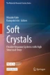
During the course of our investigations on catalytic reactions, the luminescent mechanochromism of gold(I) isocyanide complex 4 (Fig. 5.1 and 5.3) was accidentally discovered, which was reported in 2008 [16]. Pristine sample of complex 4 was a white powder and showed weak blue luminescence when irradiated with ultraviolet (UV) light (Fig. 5.3a). When a mechanical stimulus was applied to the complex, the emission of the powder changed to yellow and the maximum emission wavelength (λem,max) shifted from 415 to 533 nm (Fig. 5.3c). The single-crystal and powder X-ray diffraction structural analyses revealed that the mechanical stimulation caused a phase transition from the crystalline phase to the amorphous phase. This transformation in the crystal structure is thought to have induced a modification in the pattern of intermolecular interactions exerted on the molecules in the crystal, thereby resulting in a change in luminescence. The luminescence of gold(I) complexes has been reported to shift to longer wavelengths owing to the formation of aurophilic interactions [11, 46,47,48,49,50,51,52]. Therefore, the luminescence of complex 4 was also expected to be shifted by the formation of aurophilic interactions in the amorphous phase [16]. However, no definitive experimental data on the formation of aurophilic interactions have been reported thus far.

a Photographs (taken under UV illumination) and emission spectra of the solids of 4 before and after applying mechanical stimulation. b absorption/excitation and c emission spectra of 4 at various conditions. Adapted with permission from ref. [16] Copyright 2008 American Chemical Society, All Rights Reserved
Complex 4 was the first luminescent mechanochromic molecule reported by our research group. Isocyanide is a relatively soft ligand with a C≡N–R structure and a lone pair of electrons at the terminal carbon. Furthermore, isocyanide is known to form end-on type stable coordination complexes with various transition metals [53,54,55]. Isocyanides have a particularly high affinity for gold(I) atoms. Consequently, a variety of gold(I) isocyanide complexes have been synthesized and their physical properties have been investigated in detail [56,57,58,59,60,61,62]. Most of these gold(I) complexes form two-coordinate linear complexes. Although halogen [56], alkynyl [57,58,59,60], and cyano groups [61] have been extensively investigated as substituents on the gold(I) atom in these gold(I) isocyanide complexes, only a few examples of complexes with aryl groups as substituents on the gold(I) atom of these complexes have been reported thus far [62]. Therefore, we synthesized analogues of aryl gold(I) isocyanide complexes and successfully produced various unique mechanochromic properties, which will be discussed in the subsequent sections.
2.2 Mechanochromic Gold(I) Complexes with Tetrachromatic Luminescence
The solid sample of the diisocyanide-bridged tetrafluoropyridyl gold(I) complex 7 reversibly switches between four different color luminescences upon solvent addition and mechanical stimulation [29] (Fig. 5.4). The pristine solid sample of complex 7Y shows yellow luminescence. When 7Y was suspended in acetone, the blue luminescent phase 7B was obtained. Subsequently, the evaporation of acetone resulted in the transformation of 7B into the green luminescent phase 7G. When a mechanical stimulus was applied to 7G, the yellow luminescent 7Y was regenerated, and the application of further force resulted in its transformation to the luminescent orange powder 7O. Notably, the changes in complex 7 were found to be reversible.

Molecular structure of complex 7, photographs of the luminescent powders, and a schematic of the solid structures of each luminescent phase (indicated by rectangles and red circles for the inclusion of the solvent, acetone) and the experimental operations used to switch between luminescent phases. Adapted with permission from ref. [29] Copyright 2015 Royal Society of Chemistry, All Rights Reserved
Complexes 7B, 7G, and 7Y form different crystal structures, whereas 7O was found to be amorphous. The single crystal corresponding to 7B was obtained by recrystallization in the presence of acetone. The single crystal of 7G was obtained by the slowest possible phase transition from 7B by removing the crystallizing solvent. As for complex 7Y, it was difficult to prepare a single crystal of this complex. Therefore, the crystal structure was elucidated from powder diffraction data and Rietveld refinement. Complex 7Y is the first example of a successful ab initio structural analysis of the ground phase of mechanochromic compounds. The crystal structures of 7B, 7G, and 7Y are shown in Fig. 5.5a. In complex 7B, the intermolecular interactions between the molecules are weakened by the position of acetone (two equivalents per 7) between the molecules. In contrast, in complex 7G, the amount of solvent inclusion (acetone or water) is sufficiently small (assuming acetone, 0.5 equivalents per 7) compared with complex 7B, which allows for stronger interactions between the complex molecules (Fig. 5.5b). However, the distances between the gold(I) atoms of 7B and 7G are 3.5452(7) Å and 3.571(2) Å, respectively, which suggests that these complexes do not form aurophilic interactions. Conversely, there is no solvent inclusion in the complex 7Y crystals. The interatomic distance between gold(I) atoms is 3.428(2) Å, which confirms the formation of aurophilic interactions (Fig. 5.5c). This is thought to be the reason for the occurrence of the spectral band in the longer wavelength region compared with those of 7B and 7G. Complex 7O was identified as amorphous because no clear peaks were observed by powder X-ray diffraction.

Crystal structures of a 7B, b 7G, and c 7Y and corresponding single crystals photographed under UV irradiation. Adapted with permission from ref. [29] Copyright 2015 Royal Society of Chemistry, All Rights Reserved
2.3 Screening of 48 Complex Species for Mechanochromic Properties
A series of complexes with various substituents were systematically synthesized with the aim of controlling the emission wavelength of mechanochromic gold(I) isocyanide complexes. A total of 48 R1–R2 complexes were synthesized by introducing various substituents into the para positions of the two benzene rings, R1 and R2, using phenyl gold(I) phenyl isocyanide complex as the scaffold (Fig. 5.6). Photographs of the powdered forms of the 48 R1–R2 complex species were taken under UV light irradiation, as shown in Fig. 5.6. Each panel corresponds to an R1–R2 complex with a specific R1 substituent (vertical column) and R2 substituent (horizontal column). The substituents are arranged in order, from top to bottom and left to right, from electron-donating to electron-withdrawing groups, respectively. The pictures (taken under UV light) on the left and right of each panel correspond to the powders before and after the application of a mechanical stimulus, respectively.

Structure of R1-R2. Photographs (under UV light) of R1-R2 complexes before (left) and after (right) grinding. Adapted with permission from ref. [63] Copyright 2016 American Chemical Society, All Rights Reserved
After synthesizing the 48 R1–R2 complexes, the CF3–CN complexes were found to exhibit mechanochromism based on the crystal-to-crystal phase transition using the “screening approach” [63]. By controlling the crystallization conditions, different single crystals were successfully obtained before and after the application of a mechanical stimulus. Consequently, a clear decrease in the interatomic distance between gold(I) atoms was observed from 3.706 to 3.119 Å, which was found to be the cause of the luminescence change (Fig. 5.7). The crystal-to-crystal phase transition is relatively rare, since less than 10% of all mechanochromic molecules exhibit this phenomenon. In terms of the color of the luminescence of the R1–R2 complexes, it is found that the wavelength of luminescence color became longer as the electron-withdrawing property of the R2-position increased (from left to right in Fig. 5.6), and the luminescence color was observed to be yellow or orange instead of blue or green. Figure 5.6 shows various patterns of emission color change, and we propose that this information contained in Fig. 5.6 can serve as a library of mechanochromic molecules that can select a desired emission color change. Recently, Wu et al. reported the systematic synthesis of a variety of mechanochromic molecules by applying the C-H activation reaction to realize various luminescent colors [38].

Single crystal structure of CF3–CN species, corresponding to the molecular arrangement before (left) and after (right) CF3–CN exhibits luminescent mechanochromism. Adapted with permission from ref. [63] Copyright 2016 American Chemical Society, All Rights Reserved
2.4 Infrared Luminescent Mechanochromic Gold(I) Complexes
Extension of the π-conjugated system: Subsequently, we attempted to extend the π-conjugated system to develop a new mechanochromic molecule that emits light at longer wavelengths. The molecular structure depicted in the upper left of Fig. 5.8 corresponds to the H–H complex, as shown in the Fig. 5.6, which will be called “complex 8” hereafter. A new complex 9 was then synthesized, in which the phenyl group that binds to the gold(I) atom was extended to a naphthyl group. The emission spectrum of 9a exhibited three peaks and the λem,max was observed at 523 nm (Fig. 5.8). 9b was obtained by grinding 9a. Orange-emitting 9b had a broad emission spectrum and the λem,max was observed at 599 nm. This wavelength is longer than that of the Me-CN complex (592 nm), which exhibited the longest λem,max among the 48 R1-R2 complexes. These results indicate that the extension of the aromatic ring attached to the gold(I) atom is an effective strategy to extend the emission wavelength. Indeed, complex 10 was observed to have a λem,max in the infrared region [64].

Structures, photographs (taken under UV light), and emission spectra of complex 8, 9, and 10. Adapted with permission from ref. [64] Copyright 2017 American Chemical Society, All Rights Reserved
A novel gold(I) isocyanide complex 10, which consists of a gold(I) atom and an anthracene group, was synthesized. A pristine microcrystal of 10a exhibited blue emission. Complex 10a had a relatively sharp emission spectrum with λem,max at 448 nm (Fig. 5.8), and the absolute emission quantum yield Φem was 0.5%. The conjugated system was extended, but the emission was observed in the shorter wavelength region compared with that of 8 and 9. Notably, the average lifetime τav [= Σ(nAτ)n2/Σ(nAτn)] was found to be 0.11 ns, which was attributed to fluorescence. This result is in contrast to previously reported results of the luminescence lifetimes of aryl gold(I) isocyanide complexes, of which the majority are of microsecond phosphorescence origin.
Mechanical stimulation of complex 10a resulted in the powdered form of 10b. However, complex 10b exhibited almost no visible luminescence (Fig. 5.8). However, when the emission spectrum of 10b was measured, a broad emission was observed with a λem,max of 900 nm (Fig. 5.8) This is the longest wavelength emission observed for a mechanochromic molecule to date. Although the Φem of 10b is low (0.09%), it is reasonable to consider that the Φem values [65] of infrared-emitting solid-state organic compounds and transition-metal complexes are generally extremely low. Furthermore, the luminescence lifetime was measured to be 0.69 μs, which is typical for gold(I) complexes. The mechanochromism observed for the transformation of complex 10a to 10b shows a significant shift of luminescence to relatively longer wavelengths and a clear change in the luminescence process from fluorescence to phosphorescence. There are only a few examples [ Structure of complex 14
4.2 Gold(I) Complexes Exhibiting Optical Phase Transitions and the Photosalient Effects
Gold(I) complexes that form aurophilic interactions exhibit interesting behavior in the excited state, as shown in Fig. 5.21. As presented in Fig. 5.15, when the aurophilic interaction occurs in a pair of gold(I) complexes, the electron in the HOMO of the gold(I) complex has an antibonding character. Upon photoexcitation, the electron in the HOMO is electronically excited [Fig. 5.21(iii)]. The electrons that originally occupied the antibonding orbitals are then transferred to higher energy level orbitals, and the antibonding character between gold(I) atoms are reduced (that is, the bonding character increased). Therefore, the distance between gold(I) atoms is shorter in the excited state. This phenomenon has been investigated in detail using theoretical calculations, which has been reported [83, 84].

Schematic representation of the orbital level and the effect of the formation of the aurophilic interaction on the orbital level. Photoexcitation of a gold(I) complex that forms an aurophilic interaction typically results in an electronic transition from the antibonding dσ* orbital (HOMO) that enhances the aurophilic interaction (iii)
Although few experimental observations of enhanced aurophilic interactions upon photoexcitation have been reported thus far, Iwamura and Tahara reported results of time-resolved emission spectroscopy of a gold(I) complex [77]. The behavior of [Au(CN)2−]3 complexes in aqueous solution was investigated, and the emission of an excited species was observed at 420 nm with a lifetime of 2.0 ns. This excited species was reported to be a transient species in which the aurophilic interactions are enhanced. Furthermore, Adachi and Nozawa investigated the excited state of a gold(I) complex in aqueous solution using synchrotron radiation and reported the detailed dynamics of the formation and enhancement processes of aurophilic interactions [85]. Moreover, it is known that the intermetallic interaction of organometallic complexes with metals other than gold(I), such as platinum, rhodium, and iridium, is transiently enhanced by photoexcitation [86,87,88]. These reports confirm the enhancement of the intermetallic interaction as a transient species in solution or by measurements under UV irradiation. In contrast, we focused on the phase transition phenomenon in the crystal, and found that the gold(I) isocyanide complex 14 (Fig. 5.20) undergoes a phase transition to form a new crystal structure owing to the enhancement of the aurophilic interaction by light irradiation.
Rapid recrystallization of complex 14 in a dichloromethane-hexane solution under dark conditions yields a single crystal 14B with weak blue luminescence (Fig. 5.22, left). The absolute quantum yield Φem of the luminescence of 14B was 2.2%, and the average lifetime τav [= (ΣAiτi)/(ΣAi)] was 34.2 μs. As shown in Fig. 5.23, the emission spectrum of 14B (solid blue line) exhibits a maximum emission wavelength (λem,max) at 448 nm. The excitation spectrum of 14B monitored at 590 nm is indicated by the dotted blue line in Fig. 5.23, which had a maximum at 371 nm and showed almost no optical absorption in the visible region above 400 nm.

Single crystal structures and photomicrographs of the crystals 14B and 14Y. Excitation light used to induce the phase transition: wavelength of 367 nm, intensity of ~ 100 mW cm−2, irradiation time is indicated in the lower right corner of each photomicrograph. Excitation light for photographing: wavelength of 365 nm, intensity of < 1 mW cm−2. Adapted with permission from ref. [82] Copyright 2015 Royal Society of Chemistry, All Rights Reserved

Excitation and emission spectra of 14B and 14Y. Blue-yellow dotted lines: excitation spectra normalized to the excitation maxima of 14B (monitoring wavelength: 450 nm) and 14Y (monitoring wavelength: 590 nm). Blue-yellow solid line: emission spectra of 14B (excitation wavelength: 370 nm) and 14Y (excitation wavelength: 390 nm), which was normalized based on the intensity of the absorption spectrum. Inset: magnified view of the emission spectrum of 14Y. Adapted with permission from ref. [82] Copyright 2015 Royal Society of Chemistry, All Rights Reserved
The crystal structure of a blue-emitting single crystal 14B was analyzed by X-ray diffraction method. The space group and crystal system were determined to be P-1 and triclinic, respectively (Fig. 5.22). No residual electron density indicating solvent inclusion was observed, which was confirmed by the results of elemental analysis, thermal analysis, and 1H NMR measurements. A pair of complex 14 molecules formed a head-to-tail dimer unit. The intermolecular Au–Au distance between the molecules in the dimer was determined to be 3.5041(14) Å, which indicated that the aurophilic interactions formed were relatively weak. The dimer is then stacked to form a one-dimensional column structure, which is aligned in a sheet-like configuration with adjacent columns.
When 14B was irradiated with intense UV light, the luminescence color gradually changed, and a crystal 14Y with weak yellow luminescence was obtained (Fig. 5.22, right), after 60 s of UV irradiation. The luminescence of 14Y was very weak (Φem = 0.5%), with an λem,max of 580 nm (indicated by the solid yellow line in Fig. 5.23), which is 132 nm longer compared with that of 14B. The maximum emission wavelength of the excitation spectrum (indicated by the dotted yellow line in Fig. 5.23) also shifted by 23 nm upon light irradiation, and τav was determined to be 0.685 μs, which was approximately 50 times shorter than that of 14B.
X-ray diffraction measurements of 14Y revealed that 14B undergoes a single-crystal-to-single-crystal phase transition upon light irradiation. Several crystals of 14B were then prepared, which was successfully analyzed by single-crystal structure analysis, and irradiated using UV light under the same conditions presented above to form 14Y. Subsequently, X-ray diffraction measurement was performed. Consequently, it was reproducibly confirmed that both 14B and 14Y retained their single-crystalline nature before and after UV irradiation, even when a single piece of crystal was used. The space group of the single crystal 14Y was P-1 (Fig. 5.22). The intensity of the residual electron density was sufficiently low, which indicated that no solvent inclusion was present. Furthermore, the results of elemental, thermal and 1H NMR analyses supported this finding, which indicates that 14Y forms a head-to-tail dimer configuration with a sheet structure composed of an array of stacked column structures. These features of the crystal structure are similar to those of 14B, but differ in the following two aspects. Firstly, the 14Y molecule is significantly bent. Whereas, the gold(I) isocyanide moiety connecting the two benzene rings in the 14B molecule is nearly linear. Secondly, the interatomic distance between gold(I) atoms in 14Y was 3.2955(6) Å, which is approximately 0.21 Å shorter than that in 14B. Therefore, the aurophilic interaction was enhanced by the crystal phase transition, and caused the red shift of the emission spectrum.
DFT calculations based on single-crystal structure data revealed that the photo-induced crystalline phase transition from 14B to 14Y was induced by enhancement of aurophilic interactions in the excited state. The dimer was extracted from the single-crystal structure data of 14B and DFT calculations (PBEPBE/SDD) of this model structure were performed under various conditions. Firstly, the molecular orbitals of the 14B dimer were obtained by a single-point calculation using the crystal structure of 14B. As shown in Fig. 5.24 (i) the HOMO of the dimer has a distribution of nodes between the gold(I) atoms, which indicates that the dimer has an antibonding character. This indicates that 14B also possesses the molecular orbital features typically observed during the formation of aurophilic interactions, as shown in Figs. 5.15 and 5.21(ii). The same 14B dimer was used as the initial structure, which was optimized by assigning the overall spin as a triplet under vacuum conditions to obtain a model 14TOpt to represents the optimized structure of the triplet excited state of the 14B dimer [Fig. 5.24(ii)]. Although the head-to-tail dimer configuration was maintained, the gold(I) isocyanide unit that links benzene rings in the molecule was significantly bent. Additionally, the interatomic distance between gold(I) atoms were reduced to 2.8611 Å. These results indicate that the interatomic distance between the gold(I) atoms of the 14B dimer unit was reduced when the 14B dimer unit transitions to the triplet state upon photoexcitation. This is owing to the enhancement of aurophilic interactions by the excitation of the electron occupying the antibonding HOMO to the higher energy level orbital, as discussed earlier. Moreover, the structure of 14TOpt is similar to that of the dimer extracted from the crystal structure of 14Y [Fig. 5.24(iii), top]. These results demonstrate that photoexcitation can enhance the interaction between the gold(I) atoms in the 14B dimer, which induces the structural change to a similar structure compared with that of the 14Y dimer. These results suggest that the photoinduced crystal phase transition of complex 14B to 14Y proceeds via the rearrangement of the entire 14B molecule, owing to the attractive interactions between gold(I) atoms. To date, the photoinduced single-crystal-to-single-crystal phase transition via the enhancement of metal–metal interactions has not been reported.

Structures of the 14B, 14TOpt, and14Y dimers with diagrams of the frontier orbital energy levels and corresponding molecular orbitals. Calculation conditions: PBEPBE/SDD. Adapted with permission from ref. [82] Copyright 2015 Royal Society of Chemistry, All Rights Reserved
Furthermore, it was found that complex 14 exhibits an interesting phenomenon called the photosalient effect, in which the crystal structure changes owing to variations in the external environment, causing the crystal to “jump” (Fig. 5.25) [89]. The reason this effect is that the molecular arrangement in the crystal changes upon the application of a specific external stimulus, and strain is generated in the crystal structure. The release of this strain at a certain moment instantaneously generates a mechanical force and the crystal moves (“jumps”) into the air. The “jum**” of a crystal in response to a change in temperature is called the thermosalient effect, while that in response to light irradiation is called the photosalient effect [90]. Notably, there are fewer reports of crystals showing the photosalient effect [91,92,93,94], compared to thermosalient crystals.

Schematic of the thermosalient and photosalient effects
Complex 14 exhibited the photosalient effect when irradiated with intense UV light. When the 14B crystal was irradiated with UV light with an intensity of approximately 400 mW cm−2, the color of the luminescence changed from blue to yellow in approximately 5 s, as shown in Fig. 5.26, which indicates that the crystal phase transition from 14B to 14Y occurred. After further irradiation, small cracks appeared in the crystal, and the crystal “jumped” out of the photographic frame after 21 s (Fig. 5.26). Unfortunately, not all 14B crystals exhibited the photosalient effect under the investigated conditions (only 1% of the 14B crystals exhibited this effect). However, out of the total number of crystals tested, 80% of the crystals were cracked by UV irradiation, and 20% of the crystals were broken into two or more small pieces. In addition to the crystal “jum**” effect, a previously reported salient-active molecule also showed mechanical responses, such as cracking or breaking of the crystal surface [90]. The photosalient effect of this molecule was induced by the enhancement of aurophilic interactions by photoexcitation. In the previously reported photosalient-active molecule, the photocyclization of diarylethene and olefins recombines the covalent bonds of the molecules in the crystal, which results in a phase transition of the entire crystal, and the photosalient effect is observed [91,92,93,94]. However, the system that exhibits the salient effect without the formation or degradation of covalent bonds has not been reported thus far [82].

Photographs of the photosalient effect of complex 14. Adapted with permission from ref. [82] Copyright 2015 Royal Society of Chemistry, All Rights Reserved
Figure 5.26 shows a phase transition from 14B to 14Y before and after the photosalient effect. The features of these crystal structures are in good agreement with the typical crystal structures of previously reported salient-active molecules. Naumov and co-workers recently conducted a very intensive study on the salient effect [90]. To comprehensively discuss the mechanism of the salient effect and its relationship to molecular-level arrangements and interactions, we have thoroughly reexamined the previously reported thermosalient active molecules as well as detailed observations of the “jum**” phenomenon using single-crystal X-ray structural analysis, thermal analysis, and a high-speed camera. Based on these results, we summarize the characteristics of the crystal structures common to salient-active molecules as follows: the crystal phase transitions occurred before and after the salient effect. However, the crystal structures before and after the salient effect were very similar to each other, and the relative spatial positions of the molecules were only slightly altered. For salient-active molecules, the degree of change in the crystal structure before and after the “jump” was minimal, the space group remained the same, and the change in the V/Z values of the crystal parameters was very small (< 25 Å in all reported cases). Additionally, anisotropic changes in the crystal lattice size occurred, with a slight elongation of particular lattice lengths in certain directions and slight contraction of the other lattice lengths. Therefore, we propose that the salient effect is owing to small local strains generated by the molecular rearrangement that are released from the crystal without canceling each other as a whole. Moreover, complex 14 also satisfies many of the characteristic features of the crystal structure common to salient-active molecules, as discussed by Naumov et al. In complex 14, the space group is invariant at P-1 and the V/Z value is very small (V/Z = 6.8 Å3). Complex 14 also shows anisotropic lattice axis length variation. Therefore, complex 14 is the first case in which intermolecular interactions are essential to phase transitions in a photosalient-active molecule, and as mentioned above, it also satisfies many of the conditions for a crystal to exhibit the salient effect.
5 Conclusion
The aryl gold(I) isocyanide complexes described in detail in this paper are responsive to specific external environmental stimuli, which induce changes in luminescent color and mechanical motion. We successfully developed a mechanochromic molecule with unique features, such as multicolor and infrared luminescence properties, with the potential for various applications. Although many studies on luminescent mechanochromism have been reported thus far, the mechanochromic molecule we developed has a great advantage in terms of the characteristic features that set it apart from other studies. Furthermore, we reported a number of complexes that exhibit phase transitions between different crystals before and after mechanical stimulation or light irradiation, without loss of crystallinity, and successfully controlled the induction of luminescence changes and motions. Based on this feature, X-ray structural analysis has enabled us to elucidate the detailed molecular arrangement before and after the response, and clarify the importance of changes in the atomic interactions between gold(I) atoms. Particularly, it is noteworthy that the contrast between the type of stimulus and molecular response induced by the stimulus in the complex 8, 12, and 14. In complex 8 (and 12), mechanical stimulation induces a crystalline phase transition that strengthens (weakens) the aurophilic interactions, which resulted in a change in the luminescence properties. In contrast, in the complex 14, light irradiation induced a mechanical response in which the crystal phase transition proceeded by strengthening the aurophilic interactions, which resulted in a crystal “jump.” It can be concluded that these complexes showed stimuli-responsive properties that reflect the characteristics of the interactions formed between the gold(I) atoms. More recently, we have successfully developed a unique crystalline material that strictly deforms by the mechanical stimulation of the transition between single crystals.
