Abstract
In this study the potential of quantitative ultrasound biomicroscopy as a non-destructive methodology for the characterization of cartilage repair tissues has been investigated. In knee joints of miniature pigs, cartilage defects were treated by different cartilage repair approaches, including microfracturing, application of BMP-7, coverage with a collagen membrane, or by matrix-associated chondrocyte transplantation. After twelve weeks, the repair tissues were assessed histologically, biomechanically by indentation and by quantitative ultrasound biomicroscopy. The ultrasound signals reflected by the tissue surface and those backscattered from the internal matrix and the subchondral bone boundary were processed to estimate the integrated reflection coefficient (IRC) and apparent integrated backscatter (AIB) parameters.
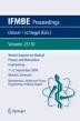
B-mode ultrasound allowed high-resolution visualization of the structure of the joint surface and subchondral bone plate, as well as determination of the thickness of healthy cartilage and repair tissues. Healthy hyaline cartilage was characterized by significantly higher IRC values and a significantly steeper negative slope of the depth-dependent backscatter amplitude AIBslope compared with the different repair cartilage tissues. Multimodal analyses revealed associations between IRC and the indentation stiffness. Furthermore, AIBslope and AIBdC were shown to be associated with the quality of the repair matrices or the reconstitution of the subchondral bone, respectively. Ultrasound biomicroscopy allowed detailed imaging of cartilage tissue, the internal tissue matrix and the subchondral bone interface. Further quantitative processing of the reflected acoustic signals contributed to a functional characterization of the biomechanical and structural properties of the cartilaginous matrix and revealed significant differences between repair tissues and healthy cartilage in this model.
Access this chapter
Tax calculation will be finalised at checkout
Purchases are for personal use only
Preview
Unable to display preview. Download preview PDF.
Author information
Authors and Affiliations
Editor information
Editors and Affiliations
Rights and permissions
Copyright information
© 2009 Springer-Verlag Berlin Heidelberg
About this paper
Cite this paper
Gelse, K. et al. (2009). Ultrasound biomicroscopy of healthy and repair cartilage tissue. In: Dössel, O., Schlegel, W.C. (eds) World Congress on Medical Physics and Biomedical Engineering, September 7 - 12, 2009, Munich, Germany. IFMBE Proceedings, vol 25/10. Springer, Berlin, Heidelberg. https://doi.org/10.1007/978-3-642-03900-3_48
Download citation
DOI: https://doi.org/10.1007/978-3-642-03900-3_48
Publisher Name: Springer, Berlin, Heidelberg
Print ISBN: 978-3-642-03899-0
Online ISBN: 978-3-642-03900-3
eBook Packages: EngineeringEngineering (R0)